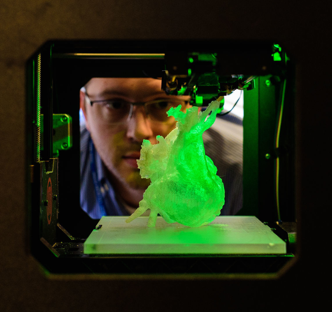
Researchers 3D bioprint realistic human heart model for the first time

Credit: Carnegie Mellon University College of Engineering
- 3D bioprinting involves using printers loaded with biocompatible materials to manufacture living or lifelike structures.
- In a recent paper, a team of engineers from Carnegie Mellon University’s College of Engineering developed a new way to 3D bioprint a realistic model of the human heart.
- The model is flexible and strong enough to be sutured, meaning it could improve the ways surgeons train for cardiac surgeries.
A team of engineers has created a new method for 3D bioprinting realistic, full-sized models of the human heart. The development could improve how surgeons train for complex procedures, and it could represent a milestone on the road toward 3D bioprinting functional human organs.
3D-printed organs aren’t a new development. But current techniques produce models that don’t feel or behave like real organs, because the printing materials are either too stiff or too soft. To create better models, Adam Feinberg, a professor of biomedical engineering at Carnegie Mellon University, and his colleagues used a technique called FRESH, or Freeform Reversible Embedding of Suspended Hydrogels.
3D-printed, lifelike heart models could help train tomorrow’s surgeons – Headline Scienceyoutu.be
The technique, described in a paper published in ACS Biomaterials Science & Engineering, uses a specialized 3D bioprinter to print soft biomaterials in a gelatin bath of hydrogel. During the printing process, the hydrogel bath helps support the delicate organ model, preventing it from collapsing. Once printed, the team applies heat to the model, causing the leftover hydrogel to melt away.
Using MRI scans of a real human heart, the team was able to 3D bioprint an accurate replica made from alginate, an affordable biomaterial that’s derived from seaweed. Alginate, which has been used in tissue engineering and wound dressing for more than a decade, has properties similar to real cardiac tissue, and it’s flexible and strong enough for surgeons to suture. That makes it an ideal material to use in training scenarios on organ models.
“We can now build a model that not only allows for visual planning, but allows for physical practice,” Feinberg said in a statement. “The surgeon can manipulate it and have it actually respond like real tissue, so that when they get into the operating site they’ve got an additional layer of realistic practice in that setting.”

Modeling incorporates imaging data into the final 3D printed object.Credit: Carnegie Mellon University College of Engineering
The FRESH technique isn’t currently able to 3D bioprint models onto which real cells can grow and form a functional heart, but similar methods may someday make that possible. If scientists can print functional human hearts, it could help the healthcare industry finally meet the demand for heart transplants, which far exceeds supply.
“While major hurdles still exist in bioprinting a full-sized functional human heart, we are proud to help establish its foundational groundwork using the FRESH platform while showing immediate applications for realistic surgical simulation,” said Eman Mirdamadi, lead author on the paper, in a statement.
In the meantime, the team behind the FRESH technique hopes to use it to generate models for other organs, like kidneys and liver.