New Brain Map Goes 2-D

What’s the Latest Development?
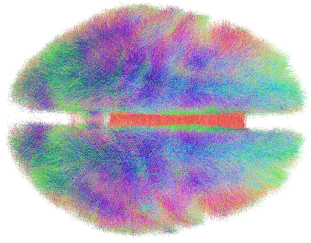
The goal of a new 2-D brain imaging technique is simplicity. Medical imaging systems that allow neurologists to summon 3-D color renditions of the brain yield valuable insights, sometimes there can be too much detail and important elements can go unnoticed. “‘In short, we have developed a new way to make 2-D diagrams that illustrate 3-D connectivity in human brains,’ said David Laidlaw, professor of computer science at Brown.” The 2-D neural maps are simplified representations of neural pathways in the brain which may give scientists a better understand of how some pathologies function in the brain.
What’s the Big Idea?
The new 2-D brain imaging process measures the water diffusion within and around nerves of the brain called myelin, a fatty membrane that wraps around axons, the threadlike extensions of neurons that make up nerve fibers. “That can help identify pathologies, such as autism, that brain scientists increasingly believe manifest themselves in myelinated axons. Diseases associated with the loss of myelin affect more than 2 million people worldwide, according to the Myelin Project, an organization dedicated to advancing myelin-related research.”


